So what type of anatomical models will help set up your physical therapy anatomy laboratory? We’ve listed a few top anatomical models used for physical therapy training and applications.
- Lifting Demonstration Figure: Provide your clients with a graphic demonstration of the effects of correct and incorrect lifting techniques on the spine.
- Shoulder Joint with Rotator Cuff 5-Part: This model shows the musculature of the rotator cuff and the origin and insertion points of the shoulder muscles.
- Knee Joint with Removable Muscles 12-Part: The 12-part knee model shows different removable muscles and muscle portions of the knee area; color coded and raised areas indicate the muscle origin and insertion points on the femur, tibia, and fibula. All the muscles of the leg are easily removable to permit study of the deeper anatomical layers.
- Functional Elbow Joint Model: This model provides an excellent graphic demonstration of the anatomy and mechanics of the joint, allowing better doctor-patient or teacher-student understanding.
- Hip Joint with Removable Muscles 7-Part: For educational purposes, the origin and insertion areas of the muscles have been raised and presented in color on the hip joint.
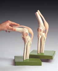
- Functional Model of the Knee Joint: In this model, the ligaments flexibility allows an excellent demonstration of the full range of motion, including flexion, extension, inner and outer rotation.
For a full list of joint anatomy models and other anatomical models for setting up your physical therapy laboratory, be sure to visit our anatomical model category on our website.


Leave a Reply